Pain that moves, appears only when you bite, or feels like sinus pressure cannot be assigned to a tooth from a scan alone. You will learn how dentists separate possible causes, choose tests and imaging that answer a defined question, and decide when a second opinion or urgent assessment matters.
Key takeaways
- Record triggers, timing, duration, and the exact area of pain.
- Expect history, examination, pulp tests, and bite checks before scanning.
- Choose imaging based on the specific question, not routine habit.
- Request copies of digital records before seeking treatment or a second opinion.
What should you record when the painful tooth is not obvious?
If you cannot identify the painful tooth, record the pattern before requesting a scan. Digital dental diagnosis for unclear symptoms in Mumbai should begin with history and examination, because pulpal pain can be poorly localised or felt in a neighbouring tooth.
A radiograph or software signal can reveal an abnormality without proving it causes your pain.
- Note when the pain began, whether it is spontaneous or triggered, and how long it lasts.
- Record the trigger: cold, heat, sweets, biting, releasing a bite, brushing, lying down or exercise.
- Mark the location and whether it shifts, spreads to the jaw, ear or temple, or feels like sinus pressure.
- Describe swelling, fever, bad taste, loose teeth, limited opening, numbness and any recent filling, injury or grinding.
- Bring previous radiographs, prescriptions and a list of medical conditions and medicines.
Seek urgent clinical care instead of waiting for digital imaging if facial or neck swelling is rapidly increasing, swallowing or breathing is difficult, you cannot open your mouth normally, or fever accompanies spreading swelling. Eye involvement or severe illness also requires prompt assessment and possible hospital care.
If findings remain unclear, the dentist can compare neighbouring teeth and use percussion, palpation, bite testing, pulp sensibility tests and periodontal probing. Sinus disease, jaw disorders and nerve pain can mimic toothache, so obtain a second opinion before root-canal treatment or extraction when the evidence does not agree.
How does a dentist narrow several possible causes?
One abnormal image does not identify the painful tooth or prove that it caused the symptom. The dentist narrows the possibilities by matching your history with examination findings, neighbouring teeth and targeted tests, then asking which diagnosis each result supports or weakens.
A typical diagnostic sequence includes:
- Visual examination for decay, cracks, leaking fillings, exposed dentine, gum changes and swelling.
- Periodontal probing to detect an isolated deep pocket that can indicate a crack or local infection.
- Bite testing to reproduce pain from a cracked tooth or a stressed filling.
- Cold, heat or electric pulp tests to measure sensory nerve response. A response shows that nerve fibres reacted; it does not prove that the pulp is healthy.
- Percussion and palpation to assess inflammation around the tooth root and surrounding tissues.
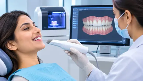
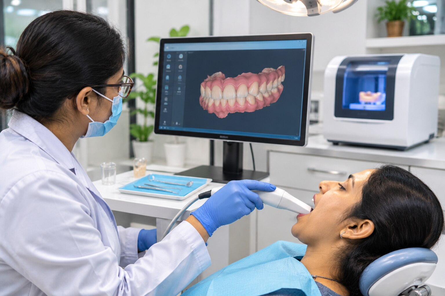
This is digital diagnosis for confusing dental symptoms only when digital findings are interpreted clinically. An intraoral scanner records the visible shape of teeth and gums as a three-dimensional surface model, including wear, tooth movement, bite contacts and restoration contours.
It cannot see inside the tooth, establish pulp vitality, reveal infection within bone or prove why a tooth hurts. A scan therefore complements, rather than replaces, examination and appropriate radiographs.
If dental findings remain inconsistent, keep non-dental causes such as sinus, jaw-joint or nerve disorders in the differential. Before root-canal treatment or extraction, seek a second opinion that reviews the original records, repeats selected tests and states what would confirm or refute each leading diagnosis.
Which imaging test answers the specific diagnostic question?
Choose the image that answers the question, not the one that produces the most data. Intraoral photographs document cracks, swelling, discolouration, gum margins and visible decay; they cannot show infection inside bone or between roots. Accurate dental diagnosis in Mumbai also depends on correct positioning, exposure, tooth labels and access to full-resolution files.
| Option | Answers best | Limits and appropriate use |
|---|---|---|
| Intraoral photographs | Visible cracks, gum changes, wear and restoration margins | Cannot assess roots, bone or hidden decay |
| Periapical radiograph | One tooth’s roots, surrounding bone, apical infection and root shape | Two-dimensional; take angled views when a fracture or extra canal is suspected |
| Bitewing radiograph | Decay between teeth and bone levels near the crowns | Does not show full root length; positioning errors can hide lesions |
| Panoramic image | Broad view of jaws, developing teeth, wisdom teeth and sinus relationships | Lower detail and distortion make it poor for small cavities or fine root fractures |
| CBCT | Three-dimensional anatomy, additional canals, resorption, cortical defects and roots near the mandibular canal or maxillary sinus | Higher radiation and metal streak artefacts; use only when simpler imaging cannot answer the question |
CBCT adds value before complex root-canal treatment, implant planning or suspected root resorption when two-dimensional views leave a specific question unanswered. It is a poor first choice for routine pain, because it can expose you to radiation without proving which tooth causes the symptom. A scan finding still requires clinical correlation, pulp tests and examination.
How can digital records improve comparison and second opinions?
Digital records improve comparison by showing what changed, where it changed and how quickly. A labelled, full-resolution intraoral scan or photograph can document tooth wear, gum contours, surface cracks, shifting teeth, bite contacts and treatment progress more reliably than memory or a compressed screenshot.
For a useful comparison, ask for:
- The original full-resolution files, not screenshots or messaging-app images
- Tooth numbers, image dates and the scan or radiograph view clearly marked
- Baseline and follow-up records displayed together
- Intraoral photographs or scans showing neighbouring teeth and the opposing bite
- Notes linking each visible change to symptoms, examination findings and treatment
A scan records visible tooth surfaces, gum margins and the three-dimensional shape of the bite. It cannot show the pulp, root or bone, and a surface line does not prove a root fracture.
Digital caries tools, including fluorescence, near-infrared transillumination and image-analysis software, can flag a suspicious area, but a positive signal does not prove cavitation or justify a filling.
Treat software output as evidence to investigate, not as the diagnosis. The dentist must correlate it with your history, visual examination, periodontal probing, bite and pulp tests, and appropriate radiographs. A second opinion is stronger when the clinician reviews those original records and explains which finding would confirm or refute each leading diagnosis.
What should you ask a clinic before accepting irreversible treatment?
Before consenting to root-canal treatment, extraction or implant placement, ask what finding proves the diagnosis and what alternative remains. If your symptoms, examination and images disagree, pause rather than accept irreversible treatment.
For technology-assisted dental diagnosis near Lower Parel, judge the process, not the scanner or CBCT brand:
1. “What is the clinical question?” Ask whether the concern is a cracked tooth, pulp disease, gum disease, bite trauma or a non-dental cause such as sinus disease or jaw-joint pain.
2. “Why is this test the lowest-risk option?” A periapical radiograph, bitewing, photograph, scan or CBCT each answers different questions; an intraoral scan records tooth surfaces and bite, not pulp inflammation or hidden root disease.
3. “Can I see the original, full-resolution images?” Check that they are labelled with the tooth, date and view, and ask whether movement, metal restorations or poor positioning limits interpretation.
4. “Who reviewed the images?” AI should support—not replace—a clinician who compares the result with examination findings and neighbouring teeth.
5. “What happens if the result remains unclear?” Ask for referral to an endodontist, periodontist, oral-medicine specialist or medical clinician when appropriate.
6. “What would a second opinion involve?” It should review original images, repeat selected tests and explain what would confirm or refute each leading diagnosis.
Whether you consult IRIDIA DENTAL or another clinic, these questions protect you from treating an abnormal image that is not causing the pain.
Related service
Technology-Driven Services We integrate digital innovation at every step—from diagnosis to treatment—to make your dental visits smoother, faster, and more precise. View service → |
Frequently asked questions
What should you record when the painful tooth is not obvious?
Record what triggers the pain, how long it lasts, whether it is spontaneous, and where you feel it. Note sensitivity to cold, heat, biting, chewing, or pressure, plus the date and frequency of episodes.
How does a dentist narrow several possible causes?
The dentist compares your history with a clinical examination, tooth-by-tooth pulp tests, percussion, bite testing, gum measurements, and checks for cracks, decay, worn fillings, or referred pain from a neighbouring tooth.
Which imaging test answers the specific diagnostic question?
A small periapical X-ray can assess roots, surrounding bone, and deep decay. Bitewing X-rays help detect decay between teeth. CBCT provides three-dimensional views when two-dimensional images cannot answer a defined question, such as complex root anatomy.
How can digital records improve comparison and second opinions?
Ask for dated radiographs, CBCT files in DICOM format, intraoral photographs, periodontal charting, test results, and the written diagnosis. A second dentist can compare the original findings rather than repeat every investigation.
What should you ask a clinic before accepting irreversible treatment?
Ask which tooth is being treated, what findings support the diagnosis, which alternatives were considered, whether observation or a reversible option is safe, and what imaging or test would change the recommendation.
Keywords